From Imaging to Simulation: The New Technical Foundation of Facelift Planning
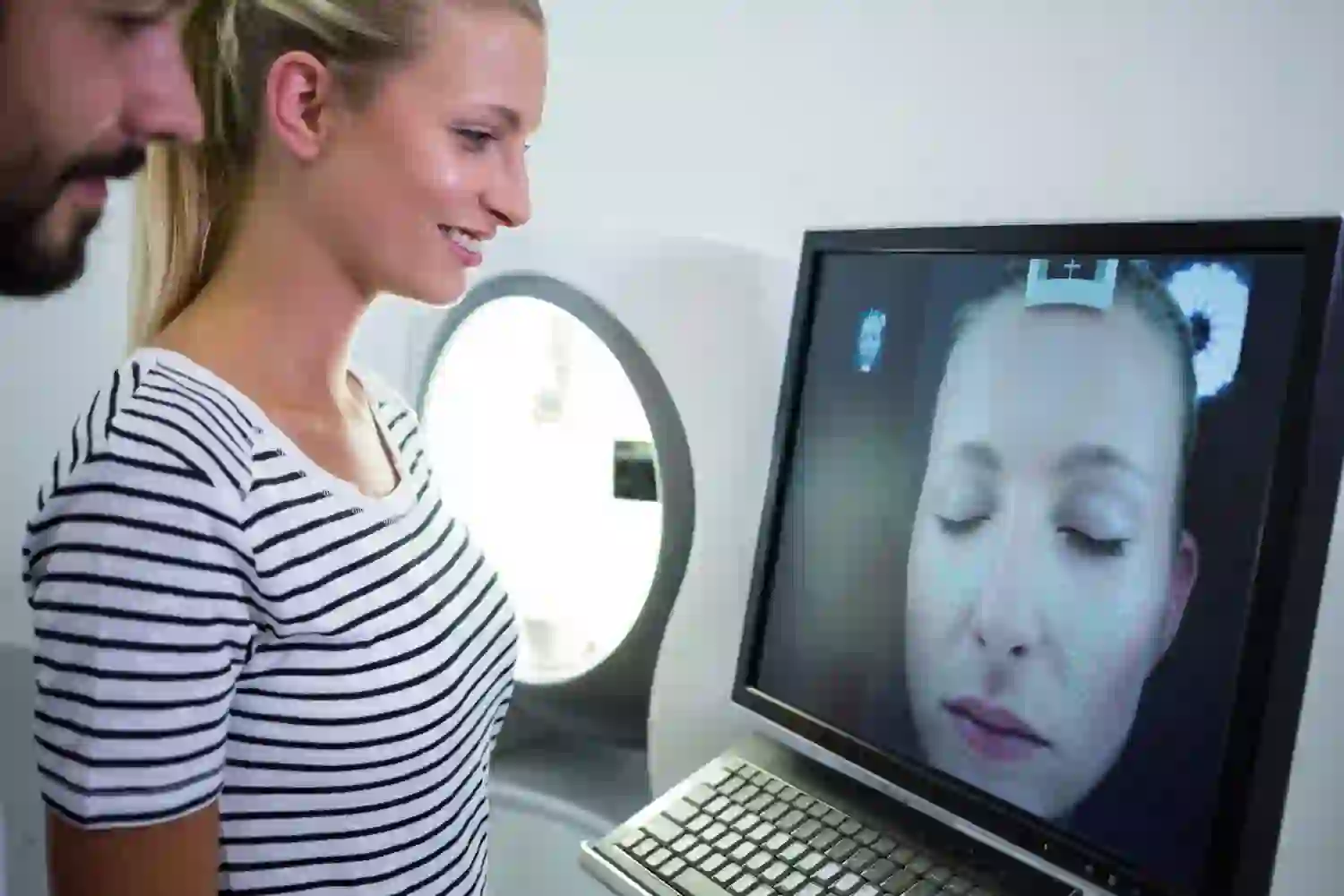
Acquisition and Registration Pipelines: 3D Photogrammetry, Structured Light, CBCT/MRI, and Landmarking
Modern planning starts with geometry you can trust. Two non-ionizing workhorses lead the way:
- 3D photogrammetry reconstructs a dense surface from multiple calibrated photos. It’s cost-effective and captures true color/texture.
- Structured light scanners project patterns onto the face and infer depth from how those patterns deform—often with higher accuracy and less sensitivity to lighting.
Need deeper anatomy? Cone-beam CT (CBCT) gives reliable bony landmarks at relatively low dose, while MRI maps soft tissue planes, parotid anatomy, and deep fat compartments—handy for complex or revision cases. Toolchains then register these modalities into a common coordinate system via:
- Rigid alignment (Procrustes, iterative closest point) to align surfaces.
- Nonrigid registration to reconcile expressions or posture shifts using free-form deformations.
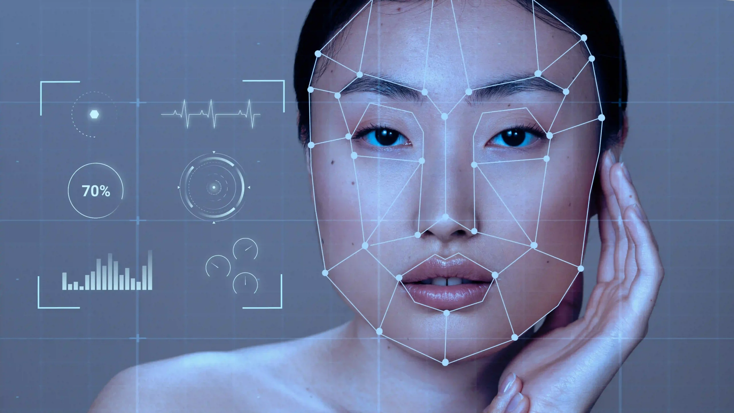
- Landmarking of perioral, periorbital, mandibular, and zygomatic points—first detected automatically by CNN-based landmarkers, then refined manually for clinical fidelity.
Well-validated pipelines can hit millimeter-level surface error under controlled conditions. That matters when a 2–3 mm change at the jowl or marionette line is clinically perceptible. Small differences, big impact.
Generative and Morphable Models for Preoperative Visualization: 3DMMs, NeRFs, and Diffusion/GAN Approaches
Once the face is registered, AI-driven generative models unlock patient-specific visualization:
- 3D Morphable Models (3DMMs) compress facial shape and albedo into low-dimensional parameters learned from large datasets. Clinicians can adjust “shape” coefficients to mimic planned maneuvers (e.g., superolateral SMAS vectoring) while preserving the patient’s identity and proportions.
- Neural Radiance Fields (NeRFs) learn a volumetric representation from multi-view images, producing photorealistic renderings under new viewpoints and lighting—great for setting expectations from every angle.
- Diffusion and GAN-based methods can synthesize textures (say, skin quality shifts) and harmonize lighting. With the right constraints, the “after” portrait matches 3D geometry—no freehand fakery.
The best systems combine morphable shape models with volumetric or neural rendering so you get clinically plausible geometry and lifelike visuals—without the “uncanny valley” of 2D edits. Because who wants a beautiful rendering that isn’t anatomically believable?
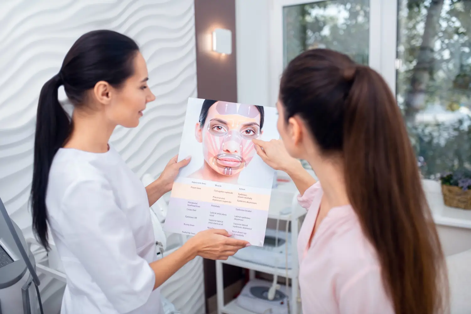
Biomechanical Soft-Tissue Modeling: FEM of Skin–SMAS–Ligament Systems and Constitutive Laws
Pretty pictures aren’t enough when skin, SMAS, and retaining ligaments respond nonlinearly to surgical forces. Finite element models (FEM) estimate how these layers deform under suture tension, undermining, release, and vectoring. Typical elements include:
- Layered structures: epidermis/dermis (anisotropic, aligned with Langer’s lines), subcutaneous fat (near-incompressible), SMAS and platysma (fiber-reinforced), and retaining ligaments (zygomatic, mandibular, masseteric).
- Boundary conditions: fixation to bony landmarks (zygoma, mandible) and deep fascia; incision edges; suture anchor points on the SMAS.
- Constitutive behavior: hyperelastic (neo-Hookean, Ogden) for soft tissue; viscoelasticity via Prony series to capture creep and relaxation; directional stiffness for collagen-heavy layers.
In practice, surgeons can run “what if” scenarios—e.g., a 20–30° superolateral SMAS vector with a target jowl displacement—then preview predicted skin redraping, nasolabial fold softening, and tension along incision lines. The goal isn’t biological perfection; it’s a patient-specific, physics-informed forecast that beats intuition alone. Helpful? Absolutely.
Validation Protocols: Surface Distance Metrics, Anthropometric Fidelity, and Intraoperative Ground Truth
Credibility demands validation:
Statistical tools like Bland–Altman plots test agreement; intra- and inter-rater reliability for landmarking ensures robustness. Clinical systems should report accuracy transparently (e.g., “1.3 ± 0.6 mm vertex-wise error over the lower face”). No smoke and mirrors—just numbers.
- Surface metrics: mean absolute surface distance, root-mean-square error, and Hausdorff distance between simulated and actual postoperative scans.
- Anthropometric checks: changes in gonial angle, lower facial third measurements, marionette fold depth, and submental plane thickness.
- Intraoperative ground truth: optical scanning of lifted flaps before closure; perfusion maps; standardized photographs for landmark verification.
Statistical tools like Bland–Altman plots test agreement; intra- and inter-rater reliability for landmarking ensures robustness. Clinical systems should report accuracy transparently (e.g., “1.3 ± 0.6 mm vertex-wise error over the lower face”). No smoke and mirrors—just numbers.
Clinical Workflow Integration: Decision Support from Consultation to Follow-Up
Preoperative Consultation and Shared Decision-Making: Scenario Simulation and Expectation Alignment
During consultation, surgeons can:
- Demonstrate multiple vectoring strategies (e.g., superolateral SMAS tightening vs. deep plane release) and show how they differently affect jowling and neck contour.
- Overlay predicted scar trajectories and camouflage strategies.
- Use outcome distributions to frame risk: “Patients with similar characteristics had X% probability of residual submental fullness at 3 months.”
Intraoperative Guidance: AR Overlays, Suture Vector Optimization, and Perfusion Monitoring Integration
In the OR, AI augments judgment—it doesn’t replace it:
- AR overlays anchored to facial landmarks can display planned incision lines, target vectors, and areas of predicted high tension. Heads-up displays or tablet-based AR keep attention where it belongs.
- Suture vector optimization tools translate planned displacements into suggested vector ranges and anchor points, based on precomputed biomechanical maps.
- Perfusion integration: real-time ICG angiography or laser speckle imaging can alert when perfusion drops below thresholds tied to delayed healing—guiding flap handling and closure strategy.
It’s guidance at the point of action, especially valuable in complex revisions or asymmetry. Small nudges, fewer surprises.
Postoperative Monitoring: Smartphone Capture, Computer Vision for Wound/Swelling, and Telehealth Protocols
Standardized at-home capture—same lighting, distance, head position—feeds computer vision models that:
- Quantify swelling volumes and color shifts as bruising resolves.
- Monitor incision lines for dehiscence risk, erythema trends, or drainage.
- Flag outliers for telehealth check-ins or earlier in-person assessment.
Automated reminders and adherence tracking keep data quality high while cutting low-value visits. Your team focuses on what really needs attention.
Interoperability and Infrastructure: DICOM/FHIR Integration, PACS Connectivity, Edge vs Cloud Compute
To avoid AI becoming a silo, make it seamless:
- Imaging: DICOM-based ingestion for CBCT/MRI; 3D photo workflows that store meshes with metadata.
- EHR: FHIR APIs to pull demographics, comorbidities, and intraoperative notes—and push structured predictions and tasks back to the chart.
- PACS/VNA connectivity for archival and enterprise access.
- Compute strategy: edge processing for low latency and privacy (e.g., OR AR overlays), cloud for heavier training and cohort analytics. A hybrid model with containerized deployments and GPU scheduling balances performance and cost.
Business Impact, Implementation Roadmap, and Future Horizons
Economic Value Proposition: Case Acceptance, Planning Efficiency, Reoperation Reduction, and KPI Frameworks
Done right, an AI platform pays its way:
- Higher case acceptance via personalized, realistic visualization.
- Planning efficiency—minutes saved per case add up fast; standardized templates reduce variability.
- Fewer revisions by de-risking asymmetries and tension hotspots.
- KPI dashboards tracking: conversion rates, planning time, revision/complication rates, time-to-edema-baseline, FACE-Q improvements, provider workload.
Change Management and Training: Clinical Adoption, Credentialing, and Cross-Functional Governance
Adoption is about people and process:
- Identify clinical champions; run simulation labs and proctored cases.
- Credential users with competency checks on capture protocols, interpreting uncertainty, and AR use.
- Stand up governance across surgery, nursing, IT, legal, and quality; define escalation paths and downtime procedures.
Build vs Buy Decisions: Vendor Due Diligence, SLAs, Validation Studies, and MLOps for Continuous Improvement
Make smart strategic choices:
Today’s decision sets tomorrow’s speed and safety.
- Vet vendors for clinical validation, data governance, security certifications, and reference sites.
- Lock in SLAs covering uptime, support, model update cadence, and issue resolution.
- Clarify data rights and IP for patient data and derived models.
- Ensure MLOps capabilities—versioning, monitoring, drift detection, A/B testing—whether in-house or via a partner.
Today’s decision sets tomorrow’s speed and safety.
Future Directions: Real-Time Intraoperative Biomechanics, Robotic Assistance, Multispectral Imaging, and Closed-Loop Systems
The next wave is already on the horizon:
- Real-time FEM approximations on GPUs to update deformation predictions as tissues are manipulated.
- Robotic assistance for steady, quantifiable retraction or suture placement under AI guidance—with force and perfusion feedback.
- Multispectral and hyperspectral imaging to map oxygenation and hemoglobin without dyes.
- Closed-loop systems that adapt intraoperative strategy based on live perfusion, tension, and predicted healing—always with human oversight.
What happens when guidance becomes truly interactive? We’re about to find out.
From Our Blog

What Is a Deep Plane Facelift and Why Do Top Surgeons Prefer It?
The deep plane facelift has become the go-to choice for many top facial plastic surgeons—and for good reason. Instead of just pulling skin tight, it works where aging actually happens: in the deeper support layers of the face.
READ THE ARTICLE
What’s the Safest Age to Get a Facelift—and When Is It Too Early?
There isn’t a magic birthday for a facelift. The “safest” age is when your anatomy and skin will clearly benefit more from surgical lifting than from nonsurgical fixes—and when your health makes an elective procedure low risk.
READ THE ARTICLE
Mini Facelift vs. Full Facelift: Technical Differences, Candidacy, and Outcomes
A facelift isn’t one single operation—it’s a range of techniques tailored to how (and where) you’re aging. “Mini facelift” and “full facelift” are common labels, but they point to very different levels of dissection, how deeply tissue is moved, and how much work is done in the neck.
READ THE ARTICLE
Is a Facelift Worth It Compared to Non-Surgical Alternatives? A Clinical, Economic, and Patient-Centered Analysis
Interest in facial rejuvenation has never been higher, but the menu of options—from injectables and energy devices to deep-plane facelifts—can feel overwhelming. Patients often ask a deceptively simple question: is a facelift “worth it” compared to non-surgical treatments?
READ THE ARTICLE