Double Board Certified · Functional Nasal Surgery
Nasal Valve Collapse — when the airway closes on inspiration.
Nasal valve collapse is a specific structural diagnosis — the narrowest part of the airway gives way on inspiration, drawing the sidewall inward and obstructing the breath. The fix is structural — and durable when done well.
ABFPRS
Facial Plastic & Reconstructive Surgery
ABOto
Otolaryngology — Head & Neck Surgery
AAFPRS
Fellowship Director
In Consultation
"Patients describe it as having to work to breathe in. The diagnosis is often missed until someone looks for it."

A Note from Dr. Mourad
"Nasal valve collapse is a specific structural diagnosis — the narrowest part of the airway gives way on inspiration, drawing the sidewall inward and obstructing the breath. The fix is structural — and durable when done well."
— Dr. Moustafa Mourad, MD
Overview
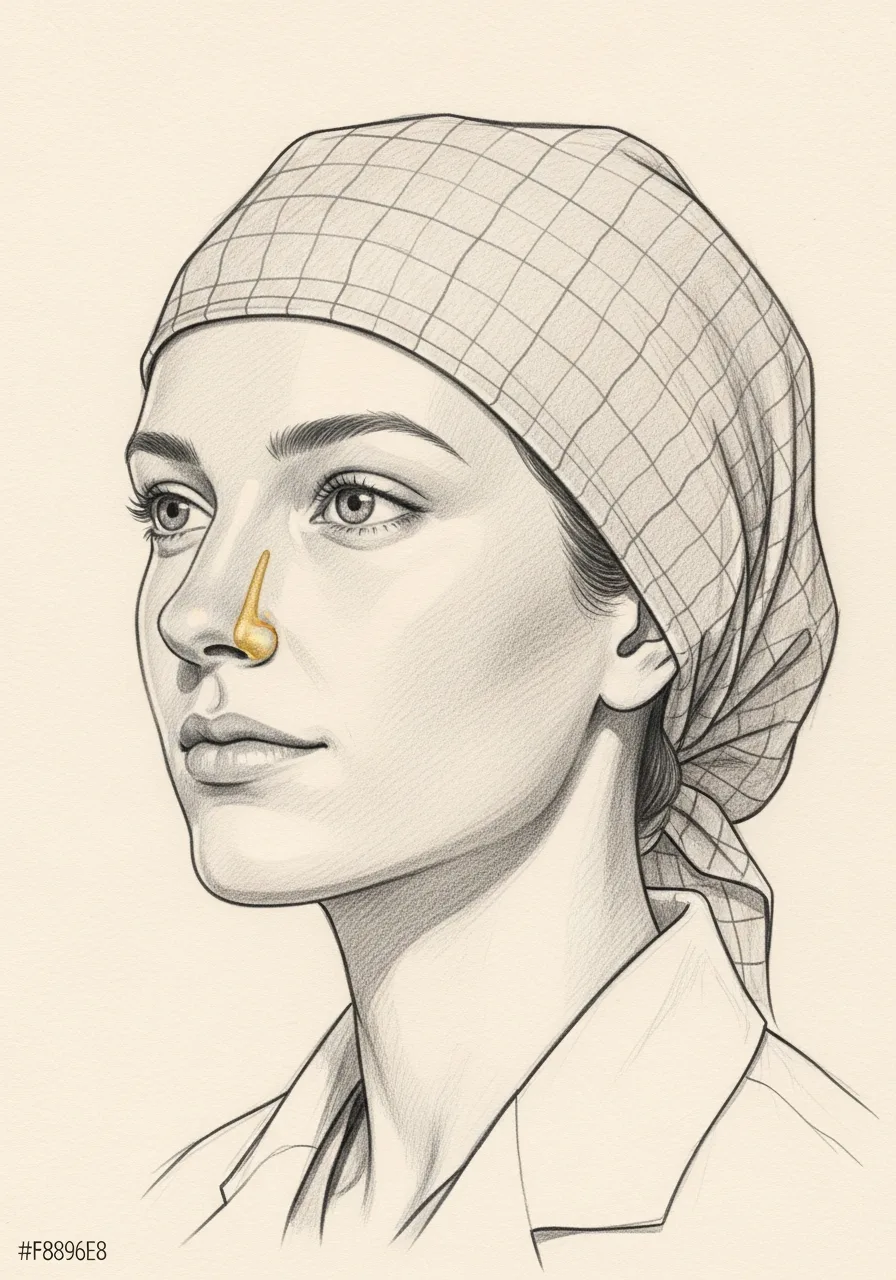
What is nasal valve collapse?
Nasal valve collapse is a structural problem in which the narrowest segments of the nasal airway — the internal nasal valve (between the upper-lateral cartilage and the septum) and/or the external nasal valve (the nostril rim and lower-lateral cartilage) — are inadequately supported and narrow or fall in during breathing.
It can be static, with the valve permanently narrow, or dynamic, with the sidewall collapsing inward on deep inspiration. Causes include congenital weakness, prior over-resection during rhinoplasty, trauma, and age-related cartilage softening. It is one of the most common under-diagnosed reasons that a septoplasty fails to relieve nasal obstruction.
Patients describe obstruction that worsens with exercise or deep breathing, an inability to wear an internal nasal strip without dramatic relief, and obstruction unrelieved by sprays or septoplasty alone. Diagnosis is made on examination, including the Cottle maneuver and dynamic visualization of the sidewall.
An Established Academic Authority
Double board certification. Fellowship director. Published author. A surgeon's surgeon.
ABFPRS
Board Certified
American Board of Facial Plastic & Reconstructive Surgery
ABOto
Board Certified
American Board of Otolaryngology — Head & Neck Surgery
AAFPRS
Fellowship Director
American Academy of Facial Plastic and Reconstructive Surgery
Textbook
Published Author
Contributions to the academic literature of facial plastic surgery
Dual board certification in both Facial Plastic & Reconstructive Surgery and Otolaryngology — Head & Neck Surgery.
Castle Connolly Top Doctor — Plastic Surgery, 202602 · Symptoms
How this condition typically presents.
Three patterns are most common. Patients often recognise themselves in one or more of these.
I
Inspiratory Obstruction
Difficulty drawing breath in through the nose, particularly with exertion. The breath out is often easier than the breath in.
II
Nasal Strip Response
Symptoms improve markedly with an external nasal strip or with a Cottle manoeuvre — a classic clinical sign of valve insufficiency.
III
After Prior Surgery
Onset or worsening after a prior rhinoplasty, septoplasty, or nasal trauma — a common pattern in revision cases.
03 · Diagnosis
How the diagnosis is made.
Diagnosis begins with a careful history — when symptoms started, what makes them better or worse, and what has been tried.
Examination includes anterior rhinoscopy and, where appropriate, nasal endoscopy with a small flexible scope to visualise the deeper nasal cavity and sinus outflow tracts.
Imaging — typically a focused sinus CT — is obtained when the examination and history warrant it, and is reviewed in detail at the visit.
04 · Treatment Options
Treatments matched to the diagnosis.
Treatment is individual. The right answer ranges from continued medical therapy to a focused minimally-invasive procedure to definitive structural surgery.
Valve Reconstruction
Structural cartilage grafts to support the internal and external nasal valves.
Learn More
Vivaer Remodeling
Minimally invasive in-office remodeling of the internal valve area for select patients.
Learn More
Revision Septorhinoplasty
When the valve insufficiency is part of a broader post-rhinoplasty problem.
Learn More
01 · Why Dr. Mourad
Diagnosis first, treatment second.
Dr. Moustafa Mourad is double board-certified in Facial Plastic & Reconstructive Surgery and in Otolaryngology — a combination uniquely suited to evaluating both the structural and the medical components of nasal and sinus disease.
Every evaluation begins with a careful history, examination, and — where indicated — endoscopy and imaging. The diagnosis is made before any treatment plan is discussed.
Medical therapy is exhausted before surgery is recommended. When surgery is the right answer, the operation is the one your anatomy and disease actually require.
When to Seek Care
When to seek care promptly.
Severe facial pain, high fever, or visual changes — these warrant urgent evaluation.
Significant facial swelling or redness around the eye — evaluate immediately.
New or worsening obstruction after a recent injury — evaluate within days.
Persistent symptoms beyond a few weeks despite over-the-counter measures — a careful evaluation is reasonable.
In Dr. Mourad's Words
Educational video.
A short educational film on nasal valve collapse from the Manhattan practice.
Nasal Valve Collapse Surgery Explained
Dr. Mourad explains nasal valve collapse — how it is diagnosed and the surgical options for restoring nasal breathing.
Outlook
What to expect.
When the diagnosis is correct and the right treatment is applied, the outlook is generally good. Many patients describe meaningful improvement in sleep, exercise tolerance, and day-to-day energy.
When symptoms persist despite treatment, the workup is re-opened. Persistent symptoms with no answer almost always mean the diagnosis is incomplete.

Living Well
Day-to-day measures that help.
Daily saline irrigation, control of indoor allergens, and good sleep hygiene meaningfully reduce day-to-day symptoms for most patients.
Medical therapy, when prescribed, works best when used consistently rather than as needed — this is one of the most common reasons treatment seems to fail.
Frequently Asked
Patient questions, honestly answered.
The nasal valve comprises the narrow internal and external regions that generate most nasal resistance. Small changes in valve angle or loss of cartilage support can markedly increase airflow resistance. Identifying whether the internal mid‑vault or the external nostril rim is failing directs treatment. A focused exam and trial mechanical tests distinguish structural obstruction from mucosal causes.
Internal valve collapse affects the mid‑vault and often becomes apparent during deep inspiration or exertion. External valve collapse shows as inward motion or pinching of the nostril rim and may be visible at rest or with inspiration. Both can coexist, and the exam uses dynamic observation plus the Cottle or modified Cottle maneuver to localize the primary site. Treatment targets the anatomic site of failure while minimizing unwanted contour change.
Candidates typically report persistent mechanical obstruction despite optimized medical therapy for at least 6–12 weeks and have a positive in‑office mechanical test such as reproducible relief with an external strip or internal dilator. Objective testing such as rhinomanometry or acoustic rhinometry can strengthen the documentation when available. Significant functional impairment—exercise limitation, sleep disturbance, or inability to tolerate CPAP—also supports consideration. Prior nasal surgery or trauma does not preclude repair but changes planning and graft selection.
Patients whose obstruction is primarily mucosal and resolves with medical therapy are unlikely to benefit from structural grafting until inflammation is controlled. Active smokers without a cessation plan, medically unstable patients, or those with unrealistic cosmetic goals are typically counseled toward optimization or non‑operative options first. Frail patients or those for whom general anesthesia is contraindicated may be offered office‑based mechanical supports or staged treatments. A consultation will clarify the best path for each patient.
Diagnosis begins with dynamic inspection and the Cottle or modified Cottle maneuver to see if lateralization improves airflow. Short diagnostic trials with external nasal strips or internal dilators are performed to reproduce symptomatic relief. When uncertainty remains, objective measures such as rhinomanometry, acoustic rhinometry, or nasal peak inspiratory flow provide quantitative data. Flexible endoscopy documents internal anatomy and excludes intranasal contributors such as polyps.
Consistent relief from nasal strips or an internal dilator indicates a structural component to your obstruction but does not automatically mandate surgery. For some patients, ongoing conservative care or a daily mechanical device is acceptable. Others with persistent, activity‑limiting symptoms after a controlled trial will likely benefit from structural support with grafting. The decision is individualized at consultation, balancing functional goals, donor‑site considerations, and aesthetic trade‑offs.
Day 0–7: expect mild to moderate congestion, drainage, and localized swelling; external splints or nasal packing are managed per procedure, and pain is usually controlled with oral medications. Week 2: external splints and many sutures are typically removed; most patients return to light work and normal sleep positioning. Weeks 3–6: swelling subsides further, moderate exercise may resume with your surgeon’s approval, and grafts begin to stabilize. Weeks 6–12: functional improvement often becomes more noticeable and contour changes settle. 6–12 months: final maturation of soft tissues and graft settling continues; if donor sites were used, their healing is usually complete. Contact the office for bleeding, persistent fever, increasing pain, or signs of infection.
Internal valve repair commonly uses spreader grafts or auto‑spreader flaps to widen the internal angle and stabilize the mid‑vault. External valve reconstruction uses alar batten grafts, lateral crural struts, rim grafts, or lateral crural repositioning to reinforce the nostril rim. Choice depends on cartilage quality, prior surgery, and whether septoplasty or turbinate reduction is required. Procedures are tailored to restore support while preserving or improving nasal contour.
Autologous cartilage—septal, conchal (auricular), and costal—remains the preferred source for long‑term support. Septal cartilage is convenient when septoplasty is performed; auricular cartilage provides thin, curved material ideal for rim grafts; costal cartilage is reserved for larger reconstructions or multiple prior surgeries. Donor‑site risks vary: septal harvest risks septal perforation or hematoma if mishandled, conchal harvest can cause contour change or pain, and costal harvest requires a chest incision and carries a rare risk of pneumothorax. Your surgeon will review donor‑site trade‑offs during planning.
Risks include bleeding or hematoma (including septal hematoma), infection, graft visibility, contour irregularity, graft warping or resorption, persistent or recurrent obstruction, and the possible need for revision surgery. Anesthesia‑related risks are present as with any operation. Many complications are uncommon; surgeons mitigate them with careful technique, appropriate graft selection, and structured postoperative care. Outcomes vary between primary and revision cases, and realistic goals are set during consultation.
Insurers typically require objective documentation of functional impairment and failed conservative therapy. Useful items include a documented trial of optimized medical therapy (commonly 6–12 weeks), reproducible relief with mechanical testing (external strip or internal dilator), physical exam findings localizing valve collapse, and objective testing when available (rhinomanometry or acoustic rhinometry). Operative reports and preoperative imaging or endoscopy that confirm structural obstruction strengthen prior‑authorization requests. Our administrative team can assist with benefit verification and submission of clinical documentation.
The Most Important Step
Get an expert evaluation.
A careful evaluation by a double board-certified physician is the right first step. The conversation is unhurried, the diagnosis is honest, and treatment is matched to what you actually have.