Double Board Certified · Revision Rhinoplasty
Pollybeak Deformity — a recognizable post-rhinoplasty problem.
A pollybeak is fullness just above the tip of the nose after rhinoplasty, creating a rounded, beak-like profile. It comes from either excess scar tissue or a structural imbalance between the bridge and tip — and the right treatment depends entirely on which.
ABFPRS
Facial Plastic & Reconstructive Surgery
ABOto
Otolaryngology — Head & Neck Surgery
AAFPRS
Fellowship Director
In Consultation
"A pollybeak is one of the most recognizable post-rhinoplasty problems — and one of the most correctable when evaluated thoughtfully."
A Note from Dr. Mourad
"Pollybeak deformity is a convex fullness in the supratip area — the region just above the tip of the nose — that creates a parrot-beak-like profile. It is most often a post-rhinoplasty problem, with both soft-tissue and structural causes."
— Dr. Moustafa Mourad, MD
Overview
What is pollybeak deformity?
Pollybeak deformity is a post-rhinoplasty contour problem in which the supratip area — the region of the bridge just above the tip — sits too high relative to the tip, producing a convex profile reminiscent of a parrot's beak. The tip can also appear under-rotated and droopy as a secondary effect.
The deformity can be structural, when too much dorsum was left or too much tip support was removed, or soft-tissue, when scar tissue forms in the supratip dead space during healing. Distinguishing the two is essential because their treatments are completely different.
Soft-tissue pollybeak may respond to steroid injections, taping, and time during the first year after rhinoplasty. Structural pollybeak requires revision rhinoplasty to rebalance the dorsum and tip. A careful examination, imaging, and review of operative records guide the choice.
An Established Academic Authority
Double board certification. Fellowship director. Published author. A surgeon's surgeon.
ABFPRS
Board Certified
American Board of Facial Plastic & Reconstructive Surgery
ABOto
Board Certified
American Board of Otolaryngology — Head & Neck Surgery
AAFPRS
Fellowship Director
American Academy of Facial Plastic and Reconstructive Surgery
Textbook
Published Author
Contributions to the academic literature of facial plastic surgery
Dual board certification in both Facial Plastic & Reconstructive Surgery and Otolaryngology — Head & Neck Surgery.
Castle Connolly Top Doctor — Plastic Surgery, 202602 · Symptoms
How this condition typically presents.
Three patterns are most common. Patients often recognize themselves in one or more of these.
I
Supratip Fullness
Visible convexity in the supratip area, most obvious on lateral profile view, creating a parrot-beak appearance.
II
Lack of Tip Definition
The tip appears under-projected relative to the supratip, blunting the natural tip-defining transition.
III
Persistent After Healing
Fullness that persists beyond the expected post-rhinoplasty swelling period — typically beyond 6 to 12 months.
03 · Anatomy
Soft-tissue vs structural pollybeak.
The treatment differs entirely. The first job at consultation is to determine which type is present.
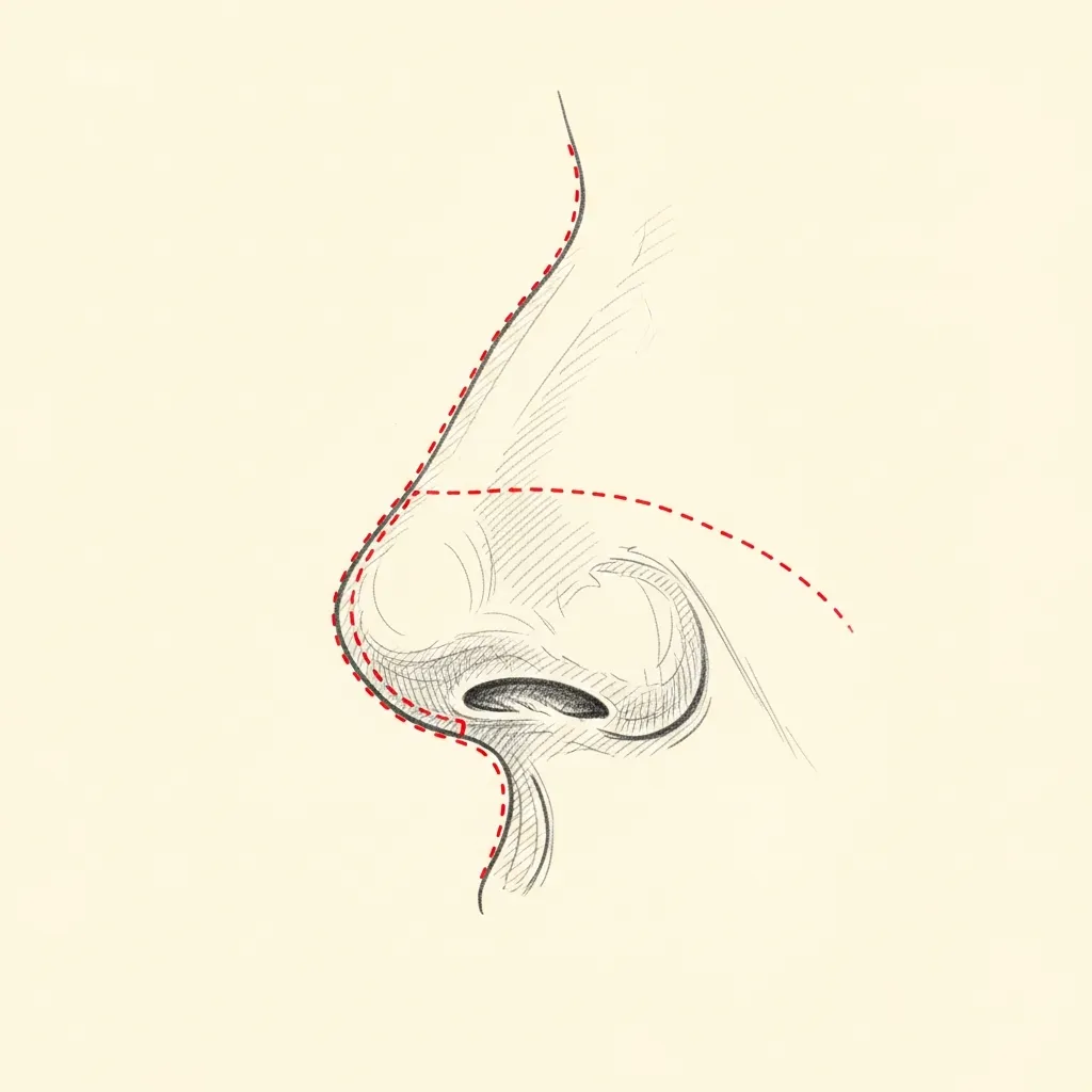
Soft-Tissue
Often responds to non-surgical management
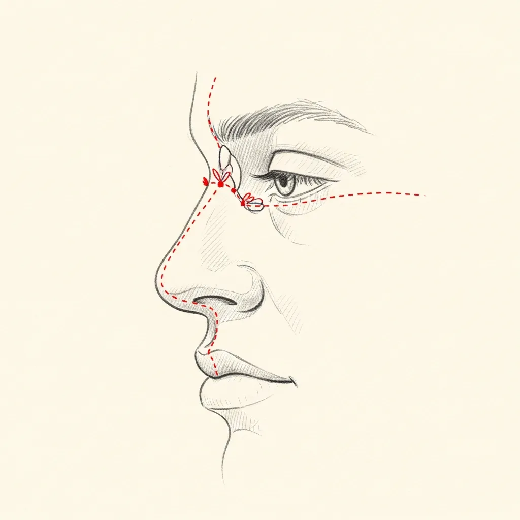
Soft-tissue pollybeak results from accumulated scar tissue and edema in the supratip area after rhinoplasty. It often improves with time and may respond to taping and selective steroid injection.
This is the more forgiving form and the right one to recognize before recommending revision surgery.
Structural
Requires revision rhinoplasty
Structural pollybeak occurs when the dorsal cartilage is not adequately reduced relative to the tip — or when the tip itself is under-projected. The bony or cartilaginous excess is visible and palpable.
Correction requires revision rhinoplasty — either reducing the supratip cartilage, increasing tip projection, or both. Revision is generally deferred at least 12 months after the prior operation.
Illustrative diagrams. The right management depends on which type of pollybeak is present.
04 · Diagnosis
How the diagnosis is made.
Diagnosis is clinical, made on examination and lateral profile photography.
The distinction between soft-tissue pollybeak (scar-tissue accumulation) and structural pollybeak (cartilage or bony excess) is the key diagnostic question.
Photographs from before and after prior surgery, when available, clarify whether the deformity is new or pre-existing.
05 · Treatment Options
Treatments matched to the diagnosis.
Treatment is individual. The right answer ranges from optimized medical therapy to a focused procedure to definitive surgery.

Non-Surgical Management
Steroid injection and taping for soft-tissue pollybeak in selected post-rhinoplasty patients.
Learn More

Revision Rhinoplasty
Structural correction for cartilage or bony pollybeak — typically requires 12 months after the prior surgery.
Learn More
Tip Refinement
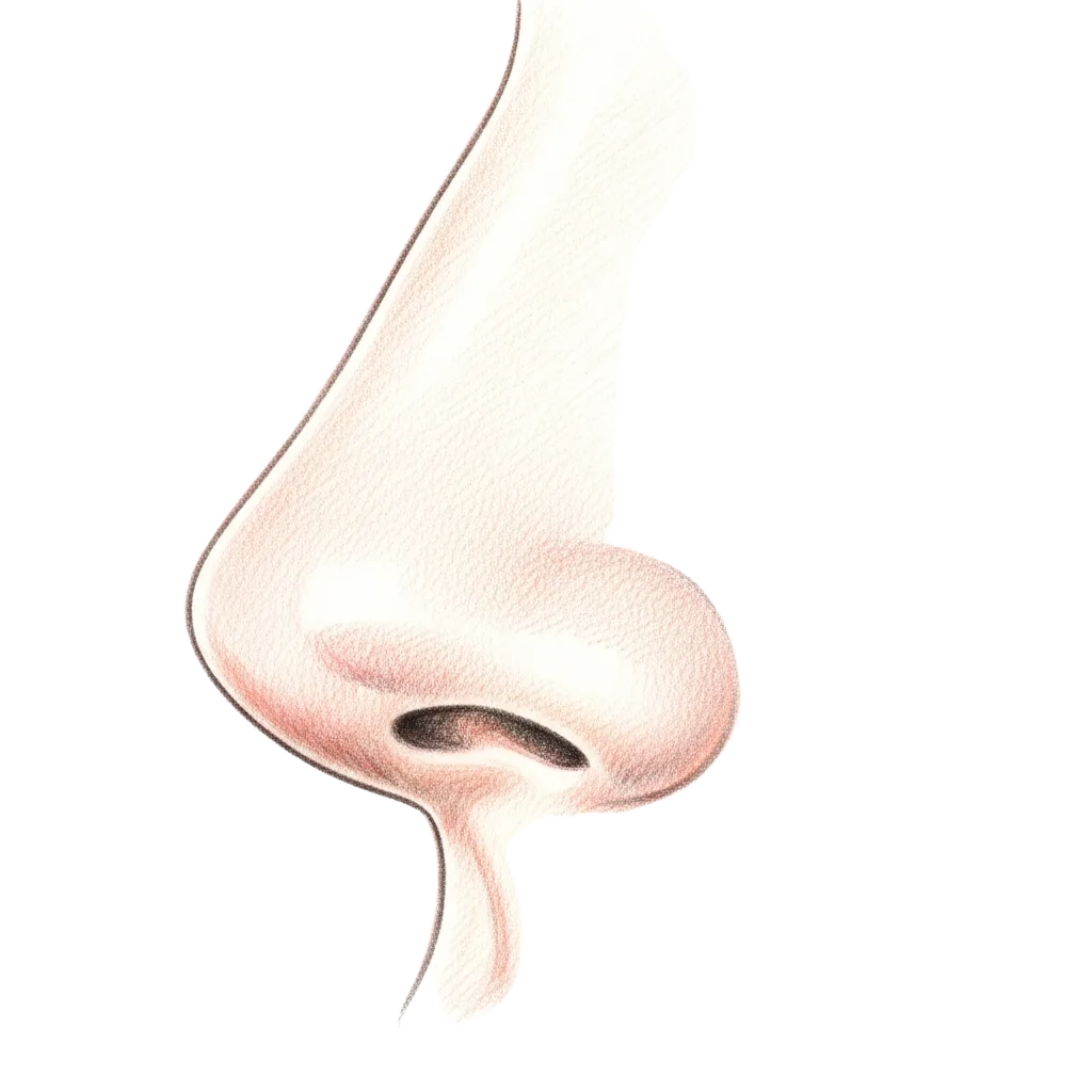
Often combined with pollybeak correction — improving tip projection is part of restoring the supratip-tip relationship.
Learn More
01 · Why Dr. Mourad
Diagnosis first, treatment second.
Dr. Mourad specializes in revision rhinoplasty and evaluates pollybeak deformity for both its soft-tissue and structural components.
A careful examination distinguishes scar-tissue pollybeak (which often responds to non-surgical management) from structural pollybeak (which requires revision surgery).
When revision is required, it is approached with the same restraint as a primary operation — the cause is corrected, no more.
When to Seek Care
When to seek care promptly.
New significant nasal asymmetry or fullness shortly after surgery — contact your surgeon.
Persistent severe pain weeks after rhinoplasty — evaluate promptly.
Sudden change in nasal shape after trauma to an operated nose — evaluate immediately.
Signs of infection (warmth, redness, drainage) after recent surgery — evaluate urgently.
Outlook
What to expect.
When the diagnosis is correct and the right treatment is applied, the outlook is generally good. Most patients describe meaningful improvement in symptoms and day-to-day function.
When symptoms persist despite treatment, the workup is re-opened. Persistent symptoms with no answer almost always mean the diagnosis is incomplete.

Living Well
Day-to-day measures that help.
Daily saline irrigation, control of indoor allergens, and good sleep hygiene meaningfully reduce day-to-day symptoms for many patients.
Medical therapy, when prescribed, works best when used consistently rather than as needed — this is one of the most common reasons treatment seems to fail.
Frequently Asked
Patient questions, honestly answered.
A pollybeak deformity is a convex supratip prominence seen on the lateral profile, producing a beak‑like silhouette above the nasal tip. Identification requires static and dynamic profile photographs and a physical exam of tip support. The surgeon differentiates soft‑tissue bulk from residual dorsal cartilage and tip ptosis. That distinction directs whether revision will focus on scar release, dorsal contouring, or structural support.
Post‑operative pollybeak commonly results from multiple interacting factors rather than a single error. Residual dorsal cartilage or intentionally preserved dorsal height can appear excessive if the tip subsequently loses support. Subcutaneous scar, skin envelope thickness, and tip ptosis from weakened support also magnify a supratip fullness. A careful review of prior operative notes, photographs, and the soft‑tissue response helps identify the dominant cause.
The Most Important Step
Get an expert evaluation.
A careful evaluation by a double board-certified physician is the right first step. The conversation is unhurried, the diagnosis is honest, and treatment is matched to what you actually have.